X-ray is an indispensable diagnostic support tool which has been used in medicine and stomatology for decades. The modern (digital) approach respecting the needs of modern dental diagnostics has become a reality in our country as well.
The advantages of digital radiology:
- Reduces radiation 50-90%.
- More precise and sharper image.
- No chemicals are used in the process of the production of the picture
- Shorter production time, by this keeping the patient
- The possibility of permanent archiving
- The carrier of X-ray images can be a film, photo paper or CD
- If the carrier is a CD the possibility of computer manipulation of the region of interest by the therapis
- Measurement of various anatomical structures in real proportions: width of the alveolar ridge, height of the alveolar ridge, length of the root canal of the teeth, the size of a tumor or cyst…
Possible techniques of filming:
The advantages of the digital radiology are obviously multiple. However, respecting the preferences and habits of a number of dentists, it can be done in the standard version on a classic X-ray film!
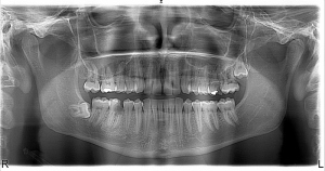
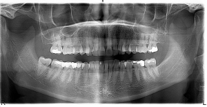
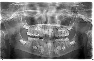
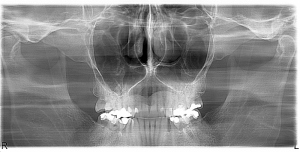
Apparatus for digital panoramic imaging methods and accurate diagnosis is the tomograph MORITA Veraviewepocs 2D.
Each patient is provided with protection against radiation of X-rays, such as impregnated clothing that is in accordance with local regulations, and appropriate infection control procedures.